X-rays, MRI, CT, Ultrasound and Scintigraphy Use in Cat Medical Care
X-rays, MRI, CT, Ultrasound and Scintigraphy – Have you ever wondered why veterinarians choose one kind of diagnostic imaging over another for your cat?
They all are a little different and can tell your vet something a little different. Depending on what’s going wrong with your cat, some are better than others.
Here are the most common diagnostic imaging tests in cats, how they work, how they are used with some advantages and disadvantages.
X-Ray (Radiographs) for Cats
How X-Ray’s Work
“You’re sending radiation through the body – ionizing radiation,” says M.C. Muhlbauer, (DVM, MS, diplomate American College of Veterinary Radiology) from Veterinary Imaging Specialists in Florida. “It goes to an imaging plate, and the imaging plate detects the radiation that went through the body. Based on how much radiation gets through, that’s how light or dark it makes the imaging plates.”
When X-rays Are Best Used
X-rays are best known for its use with bone problems and injuries, but also works well for simple contrast studies and helps veterinarians highlight possible problem areas.
Advantages of X-Rays
- Least expensive method
- Most commonly available imaging tool
- Provides a good overview of all the body’s structures
Limitations of X-Rays
“X-rays only detect 5 things: gas, fat, soft tissue or fluid, bone, and metal. You can’t see anything else. If it’s a tumor, and it’s fluid, and it’s surrounded by fluid, you’re not going to see it,” Muhlbauer says.
With general radiology, the body’s structures appear superimposed – liver on top of the gallbladder, for example. X-ray technology only gives you a brief snapshot of a cat’s anatomy. It also uses ionizing radiation which does carry some risks, less so to cats than humans as cats have a shorter lifespan than we do.
Ultrasound for Cats
How Ultrasound Works
“We’re sending noninvasive sound waves into the body and they give us real-time, dynamic imaging. With that we can see anatomy and some of the function that goes on inside the body. You can see the heart pump. You can watch the stomach contract. You can watch the intestines contract. You can watch the blood flow through the kidneys…things like that,” explains Muhlbauer.
When Ultrasound is Best Used
Ultrasound is much better than x-ray at exploring soft tissues, says Dr. Muhlbauer: “With an x-ray can see the size and shape of the liver. With ultrasound, you can actually see the blood vessels, the bile ducts, the gallbladder, the different stroma, and any masses or cysts in the liver. They all show up beautifully on ultrasound.”
Advantages of Ultrasound
- In many cases the cat does not need sedation.
- You can see the body functions, not just structure.
- Ultrasound images can be used to aid biopsies and even injectable treatments.
- You get better separation of the body’s structures which often overlap in x-rays.
Limitations of Ultrasound
- You can only see a little bit of the body at a time.
- Air blocks ultrasound, so you cannot see structures in the chest if they are surrounded by lung.
- Ultrasound cannot “see” through bone.
CT Scan for Cats
How CT Scans Works
Computed Tomography, commonly known CT scans or “Cat Scans”, are basically advanced x-rays. Dr. Muhlbauer explains: “You put the x-ray tube onto a big cylinder, and it rotates around the patient firing x-rays. On the other side of the tube is a detector. That gives us very good detail in little axial slices of the patient.”
When It’s Best Used
Subtle changes in soft tissue are captured well by CT scan technology. It’s great for looking at signs that cancer has spread to the lungs. CT scans are also good at picking up intervertebral disc disease in the spine and looking inside joints, especially elbows.
Advantages of CT Scans
- It’s faster and cheaper than an MRI.
- It typically requires less sedation than other methods.
Limitations of CT Scans
- Like x-ray, you can only see the 5 things: gas, fat, soft tissue or fluid, bone, and metal.
- Sometimes availability of this technology is a problem in smaller communities.
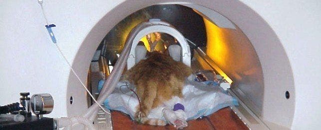
Magnetic Resonance Imaging (MRI) for Cats
How MRI Works
“Magnetic Resonance Imaging is a very powerful magnetic field that goes around the body [which] orients all the protons in your body a certain direction,” Muhlbauer explains. “As you turn off the magnets, those protons all gradually go back to their original position. [A]s they go back to their original position, they give off radio waves. There are detectors in the MRI unit that actually pick up those radio waves and use powerful computer to create an image. Depending on how fast it goes back to normal, that’s what determines the strength of the radio waves that come out, so the computer can tell what was fat, what was a tumor, what was bone, [and] what was muscle.”
When MRI Best Used
Dr. Muhlbauer explains that MRI, the “king” of neurology cases, “…can give you incredible detail, more detail than if you held the brain in your hand.” It’s also great for anything involving soft tissue and outstanding for examination of the liver, heart, kidneys, brain and spinal cord.
Advantages of MRI
- “MRI gives you the most exquisite detail,” Muhlbauer says. “If an MRI is normal, then you know there is nothing there.”
Limitations of MRI
- MRI requires sedation – often lengthy.
- It is more expensive and less available than other diagnostic imaging procedures.
- It isn’t great for imaging bones or the lungs.
Scintigraphy (Nuclear Medicine) for Cats
How Nuclear Scintigraphy Works
“With nuclear medicine, you give a radio isotope, a radioactive drug, inside the patient. Instead of radiation coming from outside and going through the patient into the detector, the radiation comes from inside the patient. The detector is called a gamma camera, and most of the radiation that comes out of these radio isotopes is gamma rays, which are exactly like x-rays but they come from a different source,” says Muhlbauer.
When Scintigraphy Best Used
Scintigraphy is thousands of times more sensitive than regular radiology. It’s great for imaging tumors and changes in bone. It provides functional, not structural, images and works well to check kidney function or blood flow patterns.
Advantages of Nuclear Medicine
- This technology is extremely sensitive.
- “It’s perfect for osteosarcoma,” Muhlbauer says. “It’ll pick up when a billionth of a gram has changed in the bone.”
Limitations of Nuclear Medicine
- It’s not very specific and can tell you something is going on, but not exactly where.
- Nuclear medicine requires expensive / special rooms and protocols for handling the radioactive isotopes.
Advances in Veterinary Imaging
I asked Muhlbauer if there were any blind spots – any places inside a cat’s body that veterinary radiologists cannot see. He replied, “I would say no.”
He then added: “I was listening to a surgeon talk the other day, and they asked him what [is] the biggest change in surgery of the last 50 years. [H]e said it was diagnostic imaging. In the past, we’d have to go inside and take a look. Now, we can use these machines to take a look inside and not have to cut the patient at all.”
So, how do veterinarians decide what imaging to use and when? Muhlbauer explains: “If an animal comes in with a complaint and needs imaging, the first thing I would do is x-ray. If I don’t get an answer with an x-ray and it’s something [related to] soft tissue, I’d go to an ultrasound….If it’s something in the lung or in bone that I cannot see, like a skull fracture, then I would next go to CT. If it were something neurological – seizures or intervertebral disc disease- I would go to MRI … I would not jump right to a CT without having x-rays first. A lot of times, x-rays give you the answer, and that saves a lot of money and a lot of stress on the patient and client.”