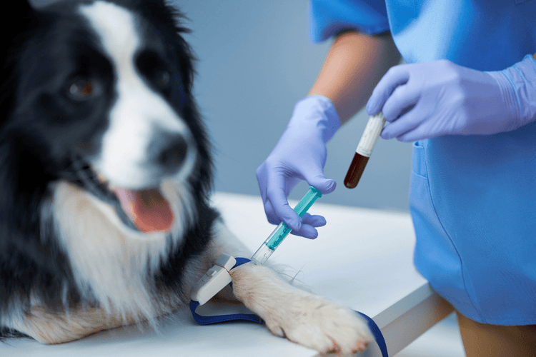
Cyanosis (Blue Coloration) in Dogs
Cyanosis is a bluish or purplish coloration imparted to the skin or mucous membranes due to excessive amounts of poorly oxygenated hemoglobin in the circulation. The causes in dogs include certain congenital heart diseases, various respiratory diseases, and exposure to certain chemicals that result in the creation of some abnormal forms of hemoglobin which are incapable of binding oxygen properly.
Cyanosis in dogs is usually an alarming clinical symptom for pet owners and for veterinarians.
Cyanosis Warning Signs
Warning signs of cyanosis include:
- Purplish/bluish coloration of the tongue, gums, lips, and areas of the skin in which the blood vessels are superficial
- Trouble or difficult breathing
- Possible purplish/bluish coloration of the foot pads
Diagnosis of Cyanosis in Dogs
A veterinarian may employ a number of methods to determine whether or not a has cyanosis and why. These include:
- Arterial blood gas measurement: Arterial blood gas (ABG) is the “gold standard” for evaluating a cyanotic patient. The test involves obtaining an arterial blood sample.
- Pulse oximetry: Pulse oximetry is readily available to most practitioners nowadays. It is a noninvasive way to get an idea of the amount of oxygen in the bloodstream. A probe is applied to a fold of skin in the axillary (armpit) or inguinal (groin) area, or the lip or tongue in an anesthetized animal
- Other specific tests, depending on the disorder that is causing the cyanosis: For example, if cardiac abnormalities are the cause of the cyanosis, cardiac ultrasound, electrocardiography, or angiocardiography may be necessary. If respiratory diseases are the cause of the cyanosis, various diagnostic tests such as thoracocentesis (removal of fluid or air from the chest cavity), a transtracheal wash, complete blood count, chemistry panel, urinalysis, chest X-rays, thoracic ultrasound, and fecal analysis may be warranted.
Treatment of Cyanosis in Dogs
Therapy of cyanosis will depend on what is causing the condition:
- Congenital Heart Disease: If the condition is caused by congenital heart disease, the treatment is surgery.
- Chemical: If a chemical has affected the hemoglobin in such a way that it cannot carry oxygen properly, for example, by inducing the formation of methemoglobin, an abnormal type of hemoglobin that cannot carry oxygen, the treatment involves: elimination of the cause, limiting any tissue injury due to poor oxygenation, and administration of medication (methylene blue; N-acetylcysteine) if necessary.
- Respiratory Disorder: If a respiratory disorder is the cause of the cyanosis, the underlying respiratory disease must be treated with antibiotics if pneumonia or chronic bronchitis is present, diuretics if fluid is building up in the lungs, thoracocentesis, which is removal of fluid or air from the chest cavity if fluid or air is causing the cyanosis, or supplemental oxygen as necessary. Emergency treatment involves making sure that the airway is unobstructed and providing oxygen.
The Two Types Cyanosis in Dogs
Cyanosis is the bluish or purplish discoloration of the mucous membranes or skin due to excessive amounts of desaturated (poorly oxygenated) hemoglobin in the blood stream. Oxygenated blood is red. Poorly oxygenated blood is dark blue. The more deoxygenated hemoglobin in the bloodstream the more bluish coloration will be imparted to the tissues.
There are two general “types” of cyanosis: central and peripheral.
Central cyanosis is a result of the entire systemic blood supply being desaturated. Central cyanosis is due to a decrease in oxygenated blood throughout the systemic circulation. All tissues are affected.
Peripheral cyanosis is due to desaturated hemoglobin that may be confined to a specific region of the body, for example, if a blood clot has obstructed blood flow to a particular body part or if a tourniquet has been applied. Peripheral cyanosis implies a purplish coloration in the peripheral tissues (oral mucous membranes, vaginal or penile mucous membranes, paw pads or nail beds, etc). All animals with central cyanosis also have peripheral cyanosis, because the entire bloodstream is desaturated. However, it is possible to have peripheral cyanosis without having central cyanosis, if the cause of the decreased oxygenation is localized to a specific region, such as a blood clot that interrupts the blood supply to a specific limb.
In young animals, the most likely cause is a congenital heart disease where poorly oxygenated blood that is returning to the heart erroneously bypasses the lungs and is sent back out into the systemic circulation without picking up more oxygen. This is called “right-to-left shunting” because poorly oxygenated blood from the right side of the heart is shunted to the left side of the heart where it is pumped out into the general circulation.
Causes of Peripheral and Central Cyanosis in Dogs
Causes of peripheral cyanosis include:
- Anything that would cause central cyanosis, with resultant bluish coloration in all peripheral tissues.
- Hypothermia. The low body temperature constricts the vessels in the skin.
- Thromboembolism, or a blood clot
- Application of a tourniquet (accidental, deliberate or malicious)
- Shock (inadequate blood flow to the tissues)
Causes of central cyanosis include:
- Congenital heart disease
- Tetralogy of Fallot, which is a genetic defect involving four abnormalities of the heart and great vessels
- Atrial septal defect (the proverbial “hole in the heart”), with subsequent right-to-left shunting
- Ventricular septal defect (“hole in the heart”) with subsequent right-to-left shunting
- Reversed patent ductus arteriosis
- Pleural effusion
- Pneumothorax
- Respiratory muscle failure
- Muscle disorder (like a diaphragmatic hernia)
- Neurologic disease
- Anesthetic overdose
- Airway obstruction
- Laryngeal paralysis
- Tumor, abscess, granuloma, foreign body obstructing a large airway
- Inadequate oxygen due to improperly administered anesthesia
- Ventilation-perfusion mismatch (improper blood supply to the lung, combined with improper lung function, or both)
- Pulmonary thromboembolism (blood clot in the lungs)
- Infiltration of the lung tissue with fluid (edema)
- Inflammatory cells (infection, inflammation)
- Cancer cells
- Acute respiratory distress syndrome (ARDS)
- Pulmonary fibrosis (pulmonary scar tissue)
Additionally, abnormal hemoglobin (methemoglobin) can result in cyanosis due to chemicals that render the hemoglobin nonfunctional:
- Nitrates
- Nitrites
- Acetaminophen (Tylenol®)
- Methylene blue
- Cetacaine
- Topical benzocaine
Emergency Measures
In cases of central cyanosis, a reduced supply of oxygen is to be assumed until it can be disproved and supplemental oxygen is to be provided until the actual cause can be ascertained. Obvious mechanical obstructions to airflow (such as a foreign body in the dog’s mouth or throat) are removed and a patent airway is established. Then, oxygen is administered immediately either by face mask, a nasal oxygen tube, an oxygen cage, or endotracheal intubation.
If congenital heart disease is the cause of cyanosis, the treatment may involve surgery to correct the defect.
If respiratory disease is the cause, the treatment is:
- Thoracocentesis to remove pus, blood, lymphatic fluid (chyle), or air that may be impeding the ability of the lungs to expand
- Antibiotics to treat infection
- Nebulization (use of a vaporizer) to moisten and loosen tenacious secretions down in the lungs and, possibly, to deliver antibiotics or other drugs down into the lungs
If excessive amounts of methemoglobin is the cause of the cyanosis, treatment involves:
- Elimination of the cause of the formation of the methemoglobin
- Acetylcysteine (Mucomyst®) can be given to dogs who have received a toxic dose of Tylenol®