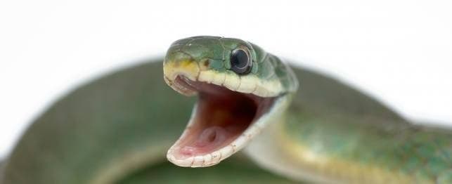
Mouth Rot (Infectious Stomatitis, Ulcerative Stomatitis)
Mouth rot is the common name used to describe mouth infections in reptiles. These infections can be of bacterial, viral, fungal or parasitic origins. Other possibilities are cancer, foreign body and jaw fractures. Poor husbandry, especially incorrect cage temperatures, poor nutrition and forced feeding predisposes reptiles to mouth infections.
Trauma to the nose or mouth areas from cage rubbing or bites from live prey are frequently associated with mouth infections. Mites and ticks often carry bacteria that can cause mouth infections, especially in snakes and lizards.
Tortoises are more prone to bacterial and viral infections that can result in mouth and lung (pneumonia) infections even when the cage environment and husbandry are excellent. If you suspect a mouth infection in your tortoise, it is always important to have it examined by a reptile veterinarian
If a lizard or snake is still eating well after consultation with your regular veterinarian, some early cases of mouth rot can be treated at home with topical medications and by improving nutrition and husbandry.
If there is significant redness, discharge, or disfigurement of the mouth or nose, or if the reptile shows a decrease in activity or appetite, it is extremely important to take it to a reptile veterinarian as soon as possible. It can be very difficult to treat mouth infections after the bone and deeper tissues are affected.
Diagnosis
Diagnosis is generally made by examining the inside of the mouth. This must be done very carefully in order to avoid trauma and tooth breakage. If an infection is not already present, a rough examination is likely to result in a mouth infection. If you are not experienced in this technique, have your reptile veterinarian examine your pet’s mouth.
Early signs of mouth rot include:
- excessive salivation
- resting with the mouth slightly open (this is also a sign of pneumonia)
- petechia, which are small red areas just under the surface of the gums where small amounts of blood are leaking out of blood vessels and they are a sign of inflammation.
Many veterinarians will want to take swabs from the mouth, wipe them on glass slides, stain them, and look at the slides under the microscope (cytology). The cytology will help determine the severity and cause of the infection. There is a blood test to check snakes for paramyxovirus and desert tortoises for some types of pneumonia that are associated with mouth infections.
As the infection progresses, the reptile often seems to be suffering from pain as he tries to eat, or he may refuse to eat at all. The petechia will become larger. Eventually , the gums will swell and except for turtles, the reptile will begin to lose teeth. At this stage, a culture of the mouth is recommended to select the correct antibiotic or antifungal medication.
In severe infections, there will be caseous exudate in the mouth, a thick, white cheesy material which is the reptile’s version of pus, and the shape of the head may become disfigured. X-rays of the head area are often required to determine the extent of bone involvement. Surgery is frequently required to remove as much of the infected material as possible and provide samples for culture or histopathology (examination of stained tissues under the microscope by a pathologist). When the infection is this severe, it often spreads to other parts of the body, so blood work to determine the body’s response to the infection and the function of organs such as the liver and kidneys is often needed.
If mouth infections are not treated early, they almost always spread to the eyes or lungs.
Treatment
Treatment of mild cases with no anorexia consists of improving the husbandry and nutrition and twice daily topical application of dilute iodine (Betadine) or chlorhexidine (Nolvasan) solution. Ask your veterinarian about the appropriate dilution for your particular pet. It is important that a solution and not a scrub is used. Scrubs contain soap and are irritating to the mouth. Hydrogen peroxide is also sometimes used as a topical medication.
In addition to the above, moderate cases of mouth rot usually require topical (applied to the mouth), parenteral (oral or injectable) antibiotics, or both. The bacteria that cause mouth infections in reptiles are often resistant to many antibiotics. Therefore, your veterinarian may need to change antibiotics once the culture results are available.
More serious cases require topical or surgical removal of the caseous debris, nutritional support (see anorexia in snakes), fluid therapy.
In mild and moderate cases, if husbandry improvements are accomplished by the owner in a timely fashion, the prognosis (estimate for getting better) for recovery is good to excellent. The prognosis is guarded for cases with significant caseous debris and grave for reptiles with significant bone involvement.
Reptiles with mouth infections should be housed at the high end of the preferred optimum temperature range. This is very important to stimulate their immune system. However, it is also important not to heat them too much because this predisposes them to thermal stress and dehydration. Ask your veterinarian for the proper temperature range for your reptile.
If you are applying topical medications or giving oral medications, make sure that your veterinarian instructs you on how to open your pets mouth safely (both for you and for your pet). Be gentle. If you are unsure, ask for more information. If you still feel uncomfortable, it may be best for your pet to be hospitalized for the topical treatments.
If your reptile eats plants, try feeding softer, less fibrous fruits and vegetables. Administer all medication according to your veterinarian’s instructions, and observe your pet’s general activity level and interest. If these worsen, contact your veterinarian.
Schedule regular veterinary visits to monitor the condition.
As a preventative measure, you should do the following:
- House reptiles at the appropriate temperature range.
- Give reptiles hide boxes to minimize cage pacing. Smooth cage surfaces minimize trauma from cage rubbing.
- Feed carnivorous reptiles previously killed prey of the appropriate type.
- Remove sticks and weeds from hay fed to tortoises.
- Control mite and tick infestations